Apple’s iTune App Store provides one standard way (and place) for “apps” developed for the iPhone and iPad to be displayed. The app store listing is really a pretty good way to learn something about an app once you’ve managed to reach the page devoted to it. Apple lets developers describe the app in under 4,000 characters and choose up to five screen shots of the app for display in its listing. The screen shots are presented without captions, so they basically need to tell their lisinopril own story.
I chose the screen shots used for the OnScreen DNA Lite listing on the iPad app store with the aim of trying to show various features, but I think a little description could be useful, so I’m presenting here those same screen shots with some explanatory text. The dimensions of these screen shots have been squeezed down to fit into the blog column, so the area of the images is less than a quarter of the iPad display’s.
Here below is the thirty-five base-pair double helix of OnScreen DNA Lite’s virtual model. Note the row of control buttons at the top. The display mode is what we have called “Balloon,” which just means that the balls used to represent molecules in the DNA structure are substantially larger than they are in the “Skeletal” mode in which the double helix structure may be more apparent. The Balloon mode is closer to the “space filling” representations sometimes shown, but not so much as to hide the structure. Since Balloon cytotec mode is in use, the button that controls this feature reads “Skeletal” to indicate that a tap of it will shift to the Skeletal representation.
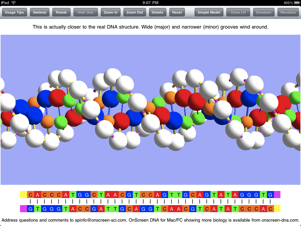
The sticks connecting the balls (molecules) in the model represent chemical bonds, which are less apparent in the Balloon mode. The model is shown above with “Tilted Bonds” (a button choice), which means that the sticks representing the glysosidic bonds between the deoxyribose phosphate molecules (white balls) and the nitrogenous bases (colored balls) are at an angle to the line between opposite sugar phosphates in the DNA strands. This propecia bond tilting is what causes the unequal spacing of the grooves (major and minor) that wind around the double helix structure. I expect to add a feature for making it obvious what these grooves are in a future update. The text in the panel above the image makes the point that the model with unequal grooves is more like the real DNA structure than the simpler model used priligy for the simulations.
Note that the bottom of the screen shots show the base sequences of the DNA strands of the model using the familiar letters GCAT (for guanine, cytosine, adenine, and thymine). The color coding is the same for the linear (letter) representation and the model.
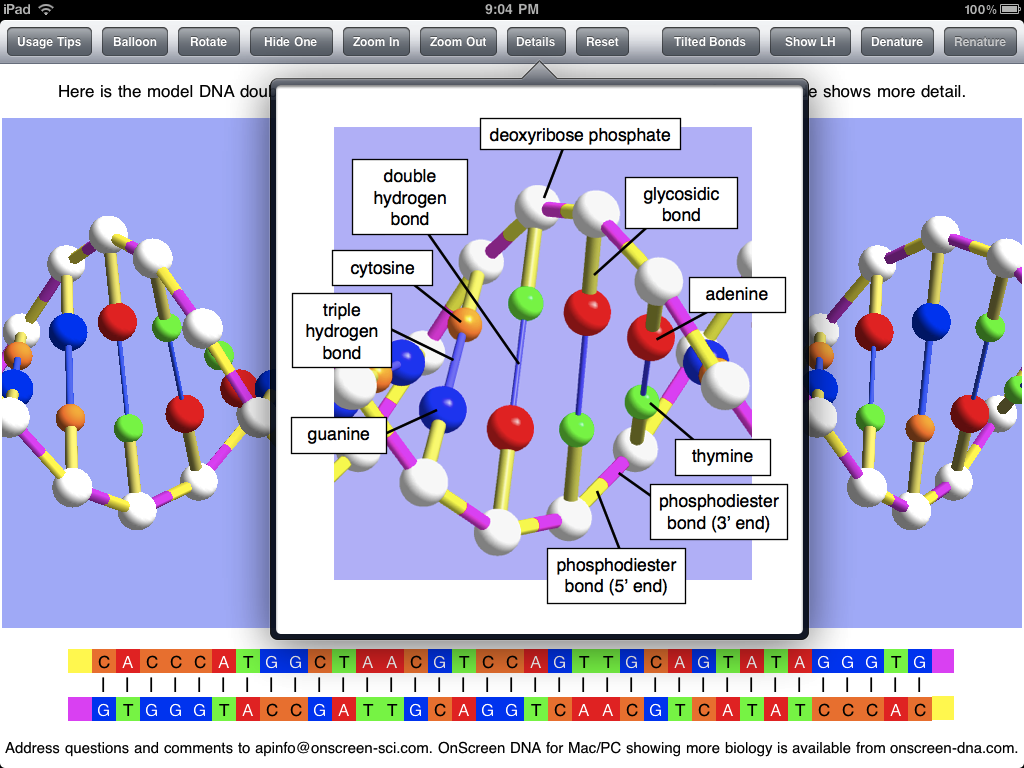
The screen shot below shows the popover view that has the key to the model of OnScreen DNA Lite. It gives the names of all the molecules and chemical bonds shown in the model. Note that the phosphodiester bond has two parts indicated. The bond is shown with two colors to make it clear that there is a polarity to the DNA strands, and that they are of opposite polarity (“point” in opposite directions).
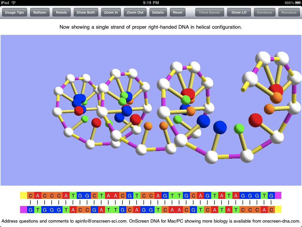
The screen shot below shows the DNA model with one of the two strands hidden, which is accomplished by a button tap. This makes the helical structure of each strand apparent. Note that this shot is with the Skeletal mode selected. Natural DNA is right-handed, meaning that a strand circles around the axis of the helix in a clockwise fashion as it advances down the axis. This handedness may be easier to see with a single strand. To further make the concept of handedness clear, OnScreen DNA Lite also has the option to show what left-handed DNA would look like. In the screen shot the model has been rotated to the side and held there. This is easily (and satisfyingly) accomplished by a swipe of a finger on the iPad screen.
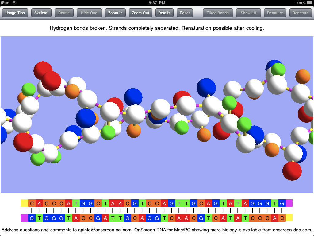
In addition to displaying the DNA model in various static (though rotatable) forms, OnScreen DNA Lite features a couple of simulations of phenomena that can occur with DNA in the laboratory. The first is denaturation, in which heating the DNA breaks the hydrogen bonds that keep the two strands joined together, thus allowing the strands to separate as single threads no longer bound to a helical shape. The screen shot below shows the two strands after denaturation has occurred, but the simulation that preceded it would have shown the strands being stretched and jiggled as the temperature increased, with individual bonds breaking until the double helix couldn’t be maintained. Note that, while showing that the hydrogen bonds are the most easily broken, an essential property for the functioning of DNA, which requires controlled strand separation at life-supporting temperatures (not the boiling temperature that brings on denaturation), denaturation is not a natural process occurring in living cells.
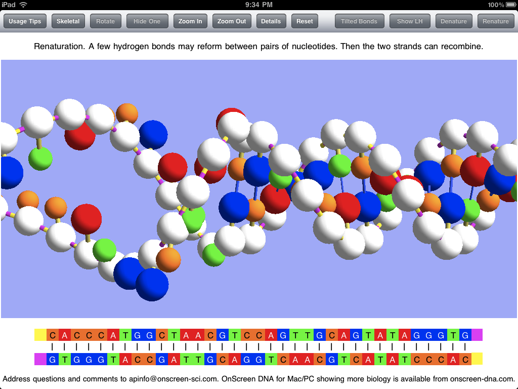
After the DNA strands have been separated in denaturation, it is possible (after the temperature has subsided) for them to recombine in the opposite process called renaturation. A few bases in one strand may come into sufficient contact with their complementary counterparts in the other strand to form a string of hydrogen bonds which can serve to hold the strands together long enough for other bonds to reform. This can be simulated in OnScreen DNA Lite after denaturation has occurred. The screen shot below captures an instant in the renaturation process after much of the double helix has been reformed, but before the process has been completed.
Screen shots can be useful in getting a picture of what an app is like, but static pictures can’t really do justice to an app with dynamic simulations and with a model that can be rotated by touch. For the true experience you’ll need an iPad and the OnScreen DNA Lite app. But soon there will be a version for the iPhone and iPod Touch. For more on OnScreen DNA Lite for iPad see The Thinking Behind the OnScreen DNA Lite™ iPad App.
Tags: DNA handedness, DNA polarity, double helix, iPad app, iPad developing, iPhone app, iPhone developing, OnScreen DNA, science simulations


 OnScreen
 OnScreen
 OnScreen