Posts Tagged ‘OnScreen DNA’

OnScreen DNA Lite™ for iPhone Now Available

Tuesday, May 11th, 2010

I’m happy to say that OnScreen DNA Lite™ for iPhone can now be downloaded from the iTunes App Store. This is basically a smaller-screen version of the iPad app that was released when the iPad first became available, though adapting the app to the iPhone and iPod Touch required some modifications, which I’ll mention. Everything I said in the blog post “The Thinking Behind the OnScreen DNA Lite™ iPad App” applies to the new iPhone version. The same desire to “provide students (and all persons interested in DNA) with a way to reach a deeper, more intuitive understanding of DNA structure” motivates the development of both apps, and the same care to show DNA’s correct handedness, base-pairs per helical turn, etc. with a ball-and-stick virtual model was taken for each. What’s more, it’s a lot of fun to play with the DNA model through the touch screen control of its orientation and size in both versions of the app.

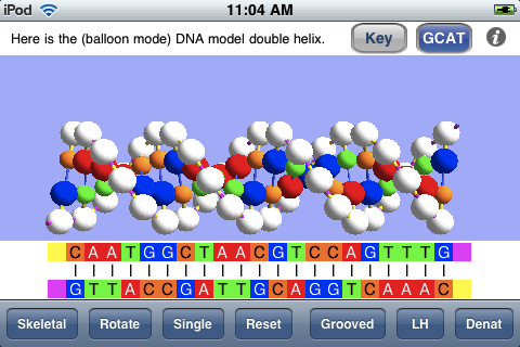
My last blog post, “An OnScreen DNA Lite™ for iPad Gallery“, showed the screen shots that are a part of the iTunes listing for that app and commented on them. As one way of comparing the two versions, let’s consider the corresponding screen shots for the iPhone app. Below is shown a screen shot from the iPhone version in which the linear (“GCAT”) representation of the base sequences of the DNA model is visible below the model. Because of the smaller screen area of the iPhone, this linear representation of the bases is only shown on demand. The button at the top designated GCAT shows and hides that view. Another accommodation to the smaller screen is the shortening of the DNA model. Instead of the thirty-five base pairs of the iPad model, the iPhone version has twenty-one, which is still sufficient to show adequately the full double helix structure and its features.

gcat

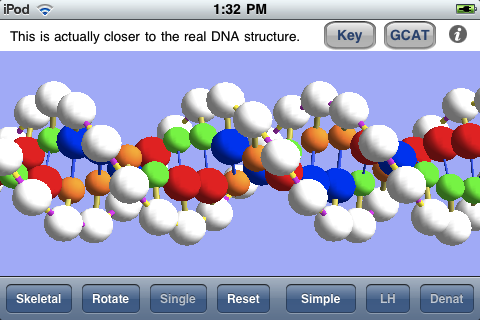
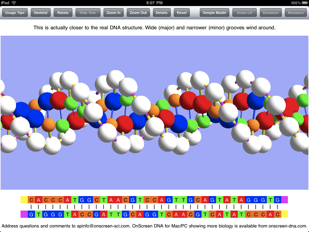
The screen shot below shows the DNA model enlarged (by means of the iPhone pinch-to-zoom technique) and with the linear view of the base sequences hidden. The structure is shown with major and minor grooves as the result of a button tap.

grooves

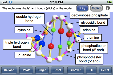
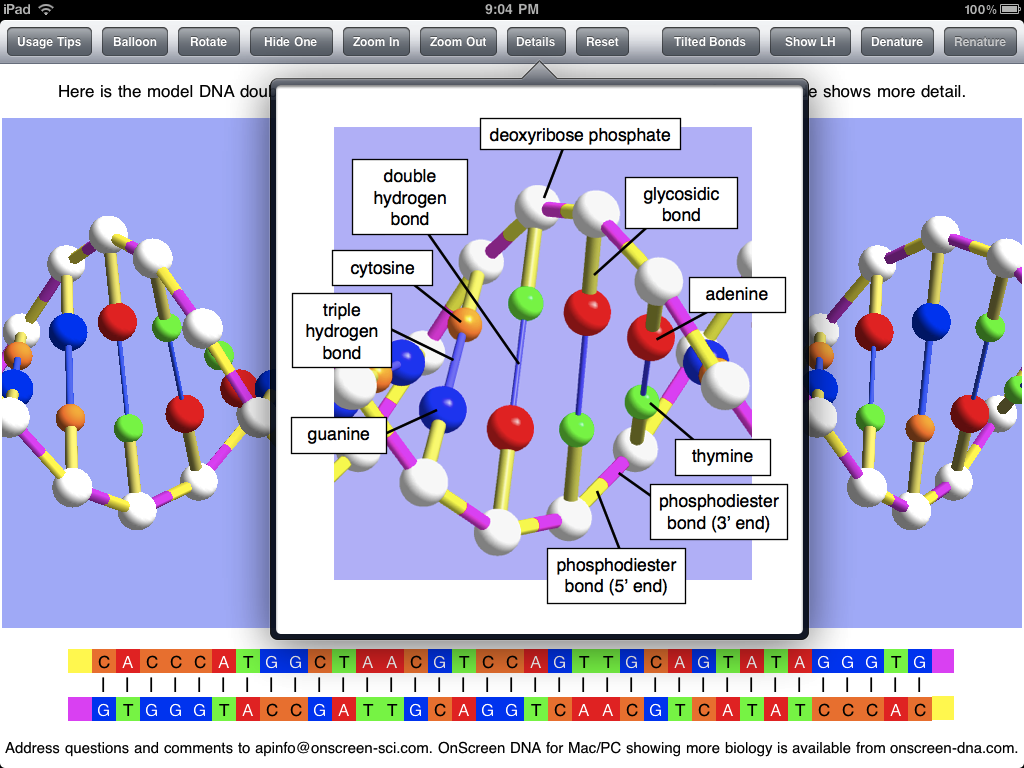
The next screen shot shows the key to the ball-and-stick model, indicating what each colored ball (molecule) and stick (chemical bond) is meant to represent. This is essentially the same view as the Details popover view in the iPad app.

key

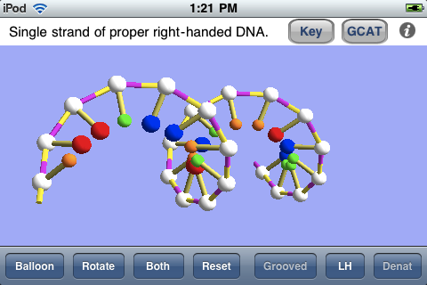
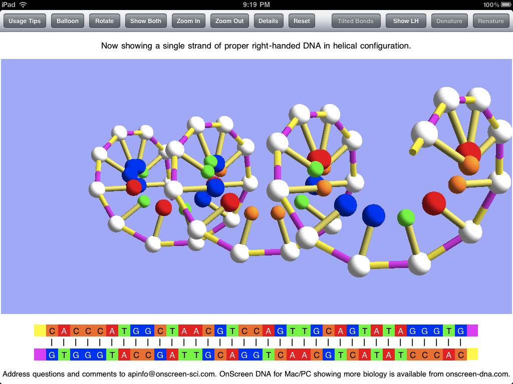
As in the iPad app, one can view a single strand of the DNA model, as shown below. This may be especially useful for grasping the meaning of the handedness of a helix, and the app also allows one to switch back and forth between natural right-handed DNA and imaginary left-handed DNA. The screen shot was taken with the model rotated by means of a finger swipe.

single

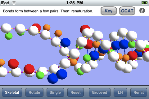
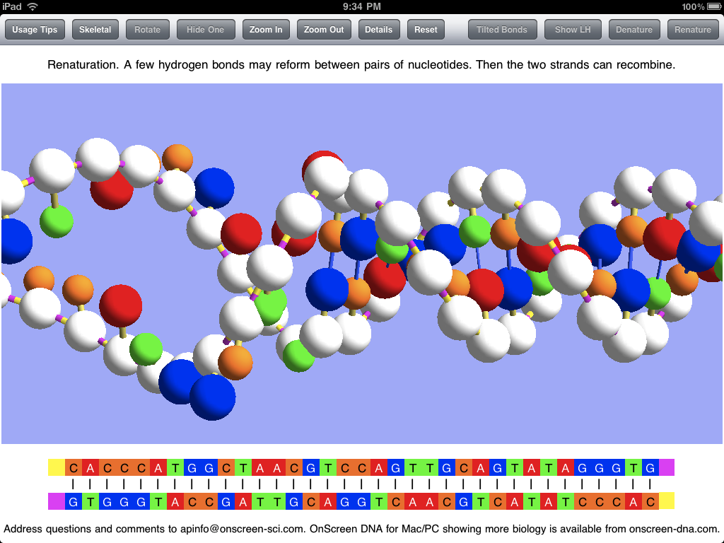
The screen shot below shows the simulated process of renaturation (rejoining together of the two strands, separated during denaturation) as it nears completion.

renat

Rather than repeat myself, I’ll just refer the interested reader to my previous two posts for more details about the virtual DNA model of OnScreen DNA Lite for iPad and now iPhone and iPod Touch.

An OnScreen DNA Lite™ for iPad Gallery

Tuesday, May 4th, 2010

Apple’s iTune App Store provides one standard way (and place) for “apps” developed for the iPhone and iPad to be displayed. The app store listing is really a pretty good way to learn something about an app once you’ve managed to reach the page devoted to it. Apple lets developers describe the app in under 4,000 characters and choose up to five screen shots of the app for display in its listing. The screen shots are presented without captions, so they basically need to tell their lisinopril own story.

I chose the screen shots used for the OnScreen DNA Lite listing on the iPad app store with the aim of trying to show various features, but I think a little description could be useful, so I’m presenting here those same screen shots with some explanatory text. The dimensions of these screen shots have been squeezed down to fit into the blog column, so the area of the images is less than a quarter of the iPad display’s.

Here below is the thirty-five base-pair double helix of OnScreen DNA Lite’s virtual model. Note the row of control buttons at the top. The display mode is what we have called “Balloon,” which just means that the balls used to represent molecules in the DNA structure are substantially larger than they are in the “Skeletal” mode in which the double helix structure may be more apparent. The Balloon mode is closer to the “space filling” representations sometimes shown, but not so much as to hide the structure. Since Balloon cytotec mode is in use, the button that controls this feature reads “Skeletal” to indicate that a tap of it will shift to the Skeletal representation.

double helix

The sticks connecting the balls (molecules) in the model represent chemical bonds, which are less apparent in the Balloon mode. The model is shown above with “Tilted Bonds” (a button choice), which means that the sticks representing the glysosidic bonds between the deoxyribose phosphate molecules (white balls) and the nitrogenous bases (colored balls) are at an angle to the line between opposite sugar phosphates in the DNA strands. This propecia bond tilting is what causes the unequal spacing of the grooves (major and minor) that wind around the double helix structure. I expect to add a feature for making it obvious what these grooves are in a future update. The text in the panel above the image makes the point that the model with unequal grooves is more like the real DNA structure than the simpler model used priligy for the simulations.

Note that the bottom of the screen shots show the base sequences of the DNA strands of the model using the familiar letters GCAT (for guanine, cytosine, adenine, and thymine). The color coding is the same for the linear (letter) representation and the model.

The screen shot below shows the popover view that has the key to the model of OnScreen DNA Lite. It gives the names of all the molecules and chemical bonds shown in the model. Note that the phosphodiester bond has two parts indicated. The bond is shown with two colors to make it clear that there is a polarity to the DNA strands, and that they are of opposite polarity (“point” in opposite directions).

popover

The screen shot below shows the DNA model with one of the two strands hidden, which is accomplished by a button tap. This makes the helical structure of each strand apparent. Note that this shot is with the Skeletal mode selected. Natural DNA is right-handed, meaning that a strand circles around the axis of the helix in a clockwise fashion as it advances down the axis. This handedness may be easier to see with a single strand. To further make the concept of handedness clear, OnScreen DNA Lite also has the option to show what left-handed DNA would look like. In the screen shot the model has been rotated to the side and held there. This is easily (and satisfyingly) accomplished by a swipe of a finger on the iPad screen.

single strand

In addition to displaying the DNA model in various static (though rotatable) forms, OnScreen DNA Lite features a couple of simulations of phenomena that can occur with DNA in the laboratory. The first is denaturation, in which heating the DNA breaks the hydrogen bonds that keep the two strands joined together, thus allowing the strands to separate as single threads no longer bound to a helical shape. The screen shot below shows the two strands after denaturation has occurred, but the simulation that preceded it would have shown the strands being stretched and jiggled as the temperature increased, with individual bonds breaking until the double helix couldn’t be maintained. Note that, while showing that the hydrogen bonds are the most easily broken, an essential property for the functioning of DNA, which requires controlled strand separation at life-supporting temperatures (not the boiling temperature that brings on denaturation), denaturation is not a natural process occurring in living cells.

denatured

After the DNA strands have been separated in denaturation, it is possible (after the temperature has subsided) for them to recombine in the opposite process called renaturation. A few bases in one strand may come into sufficient contact with their complementary counterparts in the other strand to form a string of hydrogen bonds which can serve to hold the strands together long enough for other bonds to reform. This can be simulated in OnScreen DNA Lite after denaturation has occurred. The screen shot below captures an instant in the renaturation process after much of the double helix has been reformed, but before the process has been completed.

renaturation

Screen shots can be useful in getting a picture of what an app is like, but static pictures can’t really do justice to an app with dynamic simulations and with a model that can be rotated by touch. For the true experience you’ll need an iPad and the OnScreen DNA Lite app. But soon there will be a version for the iPhone and iPod Touch. For more on OnScreen DNA Lite for iPad see The Thinking Behind the OnScreen DNA Lite™ iPad App.

The Thinking Behind the OnScreen DNA Lite™ iPad App

Friday, April 2nd, 2010

My first iPad app, now ready for sale on the iTunes App Store even before the iPad has gotten into many hands, is called OnScreen DNA Lite. Check it out! I plan to relate something of the hectic development of this app in a later post. Here my aim is to describe the iPad app a little and to motivate its development. The app is based on OnScreen DNA, a science education program I created a few years ago, first for the Macintosh OS X, and somewhat later for the Windows side. My primary goal in developing OnScreen DNA was to provide students (and all persons interested in DNA) with a way to reach a deeper, more intuitive understanding of DNA structure than I felt they were likely to obtain from reading text and looking at two-dimensional static images of a DNA model. I wanted to create a virtual, three-dimensional model that had most of the virtues of a real, physical one plus the enhanced power to simulate DNA processes with animations.

OnScreen DNA Lite’s computer model, programmed with three-dimensional perspective, is of the simple ball-and-stick type, in which the balls represent molecules, and the sticks represent the chemical bonds between these constituent molecules. A guiding principle in development was to make the relative dimensions of the model agree with those of the actual DNA molecule to the degree that makes sense for a ball and stick model. This meant getting the ratio between helical radius and the distance along the helix required for the molecular chain to make a complete revolution right, as well as showing the proper offset between molecules paired oppositely with one another in the two DNA strands. The distance between molecules in a strand, and hence the number of molecules in a complete revolution of the helical strand also had to be right.

Another crucial structural detail of the virtual helices that needed to conform with that of natural DNA was the handedness. The concept of handedness, which refers to the sense in which each helical strand winds around its axis, is one that seems largely to have escaped notice by those who make artistic renditions of DNA. My observation is that roughly half (the fraction predicted by a random guess) of all depictions of DNA show left-handed DNA, when in fact natural DNA (or all but a tiny fraction of it) in living cells is right-handed. OnScreen DNA Lite makes it easy to see the difference between right and left handed DNA by allowing the user to switch back and forth between the two.

In addition to showing the relative positions of constituent molecules in the DNA strands, the OnScreen DNA model uses color coding to identify the various molecular parts and chemical bonds. This is meant to give visual reality to the idea that a molecule of one type (color) will make a cross-strand bond with only one other type (a different color). The molecules that form a connection between their respective strands are represented by one another’s complementary colors. The color also makes the visual point that the molecules (nucleotides) making up the DNA chains differ from one another only in the parts (nitrogenous bases) that make up the cross-strand pairs, while the connections that form the individual strands are between molecular components that are identical. This can all be said, of course, and should be said, but the colors in the model make the point in an immediately memorable way.

The molecules (sugar phosphates) that link together to form the chain of a strand do so in a particular way. Think of elephants forming a line by each elephant (except for the lead elephant) grasping with its trunk the tail of the one in front of it. The molecules have an asymmetry (think of trunk and tail) as well, and they form bonds between dissimilar parts (the “tail” being the part of the molecule where the phosphorus atom is). Thus we can think of a strand of DNA as “pointing” in a given direction just as the line of elephants heads in a certain direction. We say the DNA strand has a certain polarity (as a bar magnet has polarity: N at one end, S at the other). It turns out that in the real world, the two DNA strands in a double helix are aligned with opposite polarity. They point in opposite directions. The color coding of the OnScreen DNA model reflects this feature as well, visually indicating it in the colors of the relevant chemical bonds.

In order to perform its biological function in living cells, the DNA molecule must at times have portions of its two strands separate from each other. The separation and unwinding of the strands, and the nucleic acid chain constructions involved in these processes are orchestrated by complex proteins called enzymes that catalyze just the right reactions at the right time and place in the required sequence. In the full OnScreen DNA edition, animated simulations are used to show how this occurs. OnScreen DNA Lite does not include these biological processes, but it does show how the laboratory process called denaturation takes place. The temperature required to achieve this is too high for a living cell to survive, but in the lab, the jiggling of the the double helix at the high temperarture is strong enough to break the bonds holding the two strings together. OnScreen DNA Lite for iPad animates this process, finally arriving at the point where the two strands are completely separated from each other and no longer have any helical shape, just as happens to real DNA in the lab under heating. The reverse process, in which bonds reform between complementary pairs to recombine the two strands into a double helix can also occur, and OnScreen DNA simulates this phenomenon of renaturation also.

Even though the biological functioning of DNA is not demonstrated by OnScreen DNA Lite, its animations can serve to make the point that the hydrogen bonds connecting the two intertwined strands to each other are much weaker than the other chemical bonds of the DNA molecule, a fact that is crucial for the strand separation that has to take place in the biological processes. Furthermore, I believe that seeing the strands in the act of recombining makes the fact of their entwinement all the more memorable, which is important because it seems it can be lost to consciousness when only two-dimensional images or the typical ladder-like double strand renderings are seen.

The desktop version of OnScreen DNA allows the user, by means of the mouse, to rotate the model about its helical axis and about an axis perpendicular to that. Making these rotations serves to enhance comprehension of exactly how the double helix structure is put together and to fix its three-dimensional geometrical shape in the mind. Causing the on-screen rotation by dragging the mouse pointer across the screen is fun, but the pointer on the screen is at a distance from the hand directing it. I, along with almost all other developers of iPad apps, was without the benefit of an actual iPad on which to test the app I was making and thus had to use the iPad simulator that runs on the Macintosh to see what the app should look like on the real device. Thus I was deprived of the tactile part of the iPad experience, as mouse clicks and drags had to simulate their finger-on-screen counterparts. I did, however, have a chance to test on a real iPod Touch the prototype of OnScreen DNA Lite for iPhone, and I loved how I could make the double helix rotate by moving my finger on the screen. It was much closer to dealing directly with a physical object, and much more satisfying. I can’t wait to get my iPad and to start making further improvements to OnScreen DNA Lite for iPad.

Only Three More Shopping Days Until DNA Day! Save Big!

Wednesday, April 22nd, 2009

Let’s face it: knowing the structure and workings of DNA is part of basic scientific literacy these days, which is why, after all, millions of us celebrate DNA Day. If you’re still doing last-minute DNA Day shopping, do we have a great deal for you! OnScreen DNA, the world’s best three-dimensional computer model of the double helix structure of DNA, complete with on-screen, tutorial-based simulations of how DNA works, is on sale at 50% off.

And what better way could there be to celebrate fifty-six years since the 1953 publication of the Watson and Crick paper elucidating DNA’s double-helix structure than buying OnScreen DNA for only $19.53? It’s perfect for those students, teachers, and science lovers of all ages on your shopping list. And don’t forget to treat yourself.

Don’t worry if you don’t see this until DNA Day itself—the online offer and the ability to get the software immediately by download will still be available right through April 25. Yes, we are celebrating with the “traditional” April 25 instead of moving to April 24, as many national national organizations, evidently wanting to avoid a weekend day, have done this year.

Seriously, there is nothing that I know of that teaches DNA structure and functioning in such a complete and thoroughly three-dimensional way as OnScreen DNA, which I designed and programmed myself. The software runs on Macintosh OS X or Windows XP/Vista. The on-screen tutorials explain everything you’re seeing, and practically no prior knowledge is assumed.

The animations of DNA and RNA chain-construction in OnScreen DNA are a lot of fun. I still enjoy them after having gone through them countless times during programming, debugging, testing, and just playing. You really need to see the three-dimensional structure of DNA, not just the two-dimensional ladders which animations encountered on the internet seem to invariably fall back on. Having programmed the OnScreen DNA animations, I can see why they do that—it’s a pain to do the three-dimensional programming. But it is worth it. Take a look at the results and judge for yourself. Just go to <onscreen-dna.com/buy_dna_online.php> to take advantage of this special offer.

A Commercial (with Money-Saving Coupon), Some Thank Yous, and an Animal Identification

Friday, September 26th, 2008

First, the big news: OnScreen DNA’s price has been reduced by $30! The standard edition of OnScreen DNA is now $39, and the Pro edition, which empowers user-controlled simulations of gene transcription and DNA replication, costs $69. You can read the press release; but, if you haven’t already—just to get an idea of how much easier it is to visualize and understand DNA’s double helical structure and the chemical bonds that underly it when you have a three-dimensional model to play with—why not download OnScreen DNA Lite (it’s free)?

OnScreen DNA is a virtual model, of course, which is good from a number of standpoints. It costs a lot less than a hardware one, and it can be animated to show the essential three-dimensional details of how DNA works. If you know someone who teaches DNA at any level, please tell them about OnScreen DNA. If you’ve wanted to come to a deeper understanding of DNA and how genes work yourself, please note that it is now a lot easier and less expensive to do so.

As an extra inducement to readers of this blog to try OnScreen DNA, here’s a coupon code to save an additional $20: hs908. Just enter that code in the appropriate box on the order page to get OnScreen DNA for only $19. This won’t work forever, so don’t count on it being there a month from now. OK, commercial over.

I need to catch up on thank yous and acknowledgements. As always, another blog’s linking to this one implies no endorsement of views in either direction.

David, the Christian physicist and novelist who writes the He Lives blog, linked to Conversations in the Club of Truly Smart People. Thanks again, David. Another Dave, he of the Not the Religious Type blog, mentioned the same post favorably and linked to On the Breaking of Bad Habits Acquired in One’s Youth: Smoking and Atheism. Thank you, Dave. Ropata of the Earth is My Favorite Planet blog also linked to the Bad Habits post. Thanks, Ropata.

Denyse, a very busy Catholic journalist and author on topics of religion and science, keeps three blogs going. We have exchanged some emails, and she has added the onscreen-scientist to the blog roll of Colliding Universes, which I’d say examines physics and biology from a thoughtful Intelligent Design standpoint. She also (with comments) linked to the two previously mentioned posts related to atheism and to the one on animal suffering, Cries in the Night. Thank you, Denyse.

My post about the anti-LHC campaign, Large Hadron Collider: What’s the Risk?, coming as it did a couple of days before the first proton beam circulated in the LHC, drew more traffic than even the computer troubleshooting ones have in the past. John of the Refugees from the City blog linked to my aforementioned LHC post in two separate posts: Mixed Nuts, in which he makes a thorough exposé of the dishonestly exaggerated credentials of Walter Wagner, the main instigator of the doomsday hysteria, and also looks at Rainer Plaga’s background and work, and Whooooo Hoooooo!, which summarizes the credentials of all notable LHC opponents. Thanks, John.

I have also exchanged emails with JoWynn, who wrote to tell me how much she and her husband appreciated my Reading Proust for the Last Time post. JoWynn, in addition to being a voracious reader (including books on particle physics!), maintains a blog largely devoted to her embroidery art (Parkview 616), despite a disabling condition that confines her to one room most of the time. Thanks, JoWynn. Judy of the Reading Proust in Foxborough blog said good things about the Proust post and also linked to it. Thanks again, Judy.

Finally, I’ve decided that the predatory animal whose strange wild sounds I couldn’t identify in my Cries in the Night post was almost certainly a raccoon, based on some sounds I’ve found online. It’s funny that out of all the raccoons I’ve seen in my life, I’ve never heard one make a sound that I can remember. So, just to return to that disturbing death struggle I overheard in the middle of the night, I now imagine that it was a raccoon that had caught a squirrel. The raccoon, lacking big, powerful jaws like a dog, could have been holding the squirrel in its mouth waiting for it to die of blood loss, internal injuries, etc. The squirrel, being still alive, could have made its cries and also have mustered up the strength for a desperate struggle to escape every few minutes, which would explain the fierce raccoon sounds mixed with thrashing around that I heard periodically.

On the one hand, I’d just as soon get those sounds and speculation about what was going on out of my head, but it’s also good to have the drama linked to known animals. It changes my view of raccoons, which I had known to be scrappy fighters by reputation (able to drown dogs that were foolish enough to pursue them into the water, for example), but had never seen or heard in action.

Commercial Break: DNA Day Sale Now Underway

Friday, April 18th, 2008

And now, a word from our sponsor. If you’ve ever wanted to learn or thought that maybe you should learn what the structure of DNA really looks like and how DNA actually works in our cells to make us who we are and keep us going, then you have a chance to do it in a very thorough and enjoyable way for the lowest price ever. OnScreen DNA, the virtual model programmed by the On-Screen Scientist himself, is on sale at a 70% markdown, in honor of DNA Day and the original discovery of the DNA double helix fiftty-five years ago. The price? Just $19.53 (where’d that number come from?), instead of the usual $69, through April 25, 2008.

What’s DNA Day? It’s the day we celebrate both the historic publication of the Watson and Crick paper that elucidated the double helical structure of DNA and the completion of the Human Genome Project. April 25 has been chosen as the day. I may feel moved to say something about the discovery one of these days, maybe even before DNA Day, but for now I wanted to alert my many readers to the sale.

The software runs on Macintosh OS X or Windows XP/Vista. Download the free OnScreen DNA Lite first if you wish to see the quality of the model. But you don’t really get the full how-DNA-works story in the Lite version, just a detailed guided tour of the structure, including the essential molecular components and chemical bonds. The on-screen tutorials explain everything you’re seeing, and practically no prior knowledge is assumed.

Can you spot the many (roughly 50% it seems) inaccurate popular depictions of DNA as a left-handed helix instead of the proper right-handed type? Well, you’ll be able to after a few minutes with OnScreen DNA (or even the Lite edition, for that matter). It would be so easy to tell the artists hired to make all these nifty DNA double helix logos and designs the difference between left-handed and right-handed DNA, but it seems no one does, even scientists. Witness the many backward examples. I think the professor that was maintaining the left-handed-DNA web site finally got tired of it, but I imagine the archives are still up in any case. Just Google it. New examples appear all the time. The Boston Globe had a doozy that occupied about half a page just last week. Left-handed DNA does exist in nature, but it’s a small percentage and is not the genome molecule of our chromosomes.

Anyway, the animations of DNA and RNA chain-construction in OnScreen DNA are a lot of fun. I still enjoy them after having seen them countless times during programming, debugging, testing, and just playing around. You really need to see the three-dimensional structure of DNA, not just the two-dimensional ladders which animations encountered on the internet seem to invariably fall back on. Having programmed the OnScreen DNA animations, I can see why. It’s a pain to do the three-dimensional programming. But it is worth it. Just go to <onscreen-dna.com/buy_dna_online.php> to purchase online and see for yourself. Or get the free version from the link in the upper right. Amaze your friends by pointing out the next picture of left-handed DNA you come across! That’s almost as good as ordering in French at the French restaurant.